Skelettröntgen
Application
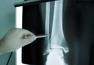
As a rule, skeletal x-rays of joints and bones are taken in two planes, often with the patient recumbent but in some instances erect, when there are particular issues.
In very rare instances, it is also necessary to take images from the opposite side for purposes of comparison. Depending on the region examined, you will be asked to remove items of clothing and jewellery.
Indications
- Assessment of sections of the skeleton after accidents, detection of potential fractures
- Signs of wear in joints (arthrosis)
- Changes in bone structure (osteoporosis, Paget’s disease)
- Check on an artificial joint (Please bring previous images!)
- Check on osteosythesis material (DHS, Gamma nail, intramedullary nail, PLIF, Harrington rods – please bring previous images!)
- Inflammatory changes in bones (osteomyelitis)
- Inflammatory joint changes (arthritis)
- Benign and malignant tumours
- Malposition or defective position (scoliosis)
- Inflammation and air-fluid level in the paranasal sinuses
- Bone-age assessment